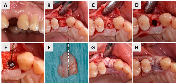
Figure 7.
Implant placement and soft tissue augmentation. (A) Buccal view of the surgical site at tooth 24 (FDI Notation) after six months of healing. The stable contour of the buccal soft tissue and preserved alveolar ridge are evident; (B) Elevation of the full-thickness mucoperiosteal flap, exposing the underlying alveolar bone for implant bed preparation; (C) Bone biopsy being obtained using a trephine bur with a 2 mm inner diameter, which was carefully chosen to avoid damage to the implant bed; (D) View of the implant bed; (E) Placement of the dental implant (4.1 × 10 Straumann® BLT, Basel, Switzerland) into the prepared implant bed. The probe revealed a sufficient buccal bone wall thickness of approximately 3 mm surrounding the implant; (F) A free gingival graft, harvested from the maxillary tuberosity and deepithelialized extraorally, was prepared for augmentation to enhance the soft tissue volume around the implant site; (G) The tissue graft was secured over the buccal aspect of the implant with 6-0 sutures (Surgicryl® Monofast, St. Vith, Belgium) to enhance tissue thickness and promote optimal healing; (H) Primary wound closure was achieved with interrupted 6-0 non-resorbable sutures, ensuring tension-free closure and promoting uneventful healing.

