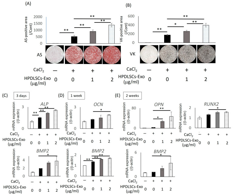
Figure 3.
Effects of HPDLSCs-Exo on differentiation of Saos2 cells. Saos2 cells were cultured in α-MEM containing 10% exosome-depleted FBS (control), control containing 1 mM CaCl2 (Ca), and Ca with 1 or 2 µg/mL of HPDLSCs-Exo. (A,B) Alizarin red S staining (AS) and von Kossa staining (VK) were performed after 2 weeks of culture. AS-positive areas (A) and VK-positive areas (B) were quantified. * p < 0.05, ** p < 0.01, n = 3. (C–E) Saos2 cells were cultured in control, Ca, and Ca with 1 or 2 µg/mL of HPDLSCs-Exo for 3 days (C), 1 week (D), and 2 weeks (E). Quantitative RT-PCR was performed to analyze the expression of bone-related markers such as alkaline phosphatase (ALP), bone morphogenetic protein 2 (BMP2), osteocalcin (OCN), osteopontin (OPN), and runt-related transcription factor 2 (RUNX2). Normalization of gene expression was performed against β-actin expression, and the gene expression levels were shown as the fold increase relative to controls. * p < 0.05, ** p < 0.01, n = 3.

