Figure 5.
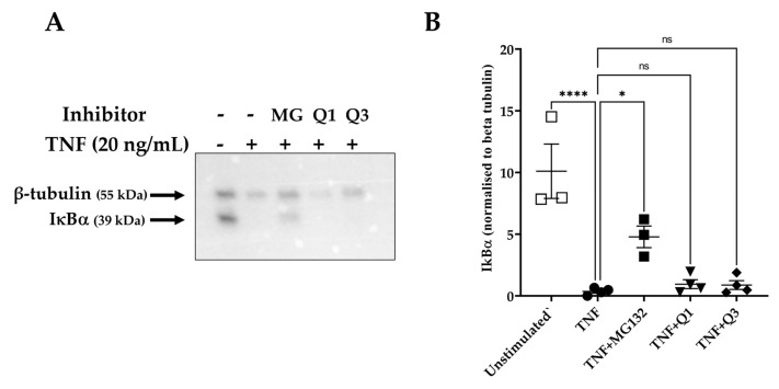
IκBα degradation as assessed by immunoblot analysis in TNF-induced HeLa/NF-κB-Luc reporter cells. HeLa/NF-κB-Luc cells were either left unstimulated (n = 3) or stimulated with 20 ng/mL TNF (n = 4) for 20 min in the absence or presence of 40 μM MG132 (MG) and 10 μM Q1 or Q3 (all n = 4). (A) RIPA cell lysates were analyzed with SDS-PAGE, transferred to a PVDF membrane, and levels of IκBα and β-tubulin were each detected by an immunoblot analysis using specific antibodies. Immunoblots are representative of four experiments. All immunoblot experiments can be reviewed in Supplementary Information File S4. (B) A quantification analysis of the IκBα bands normalized against β-tubulin levels was performed. One-way ANOVA followed by Tukey’s post hoc multiple comparisons test revealed significant differences. * p < 0.05, **** p < 0.0001. Non-significant, ns.

