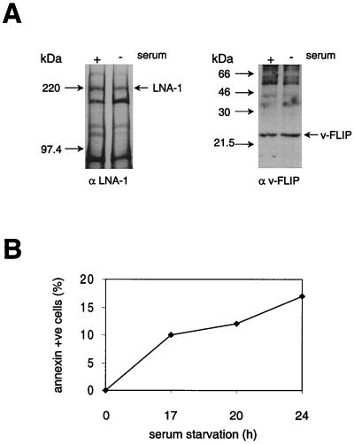
FIG. 7.
Synthesis of LNA-1 and v-FLIP during apoptosis of PEL cells. BC3 cells were serum starved for 20 h to induce apoptosis, which was detected by ANNEXIN-V Fluos staining (B) as described in Materials and Methods. Viable cells (107) were then pulsed for 2 h with labeled methionine and cysteine, after which LNA-1 and v-FLIP were immunoprecipitated as described in Materials and Methods and the immunoprecipitates were separated by SDS–7 or 14% PAGE, respectively (A). The relative levels of labeled protein were determined using densitometry software on a phosphorimager as described in Materials and Methods. The FLIP/LNA-1 labeled-protein ratio was normalized to 1 in cells with serum; the relative FLIP/LNA-1 labeled-protein ratio in cells without serum was 3.25. In a second experiment cells were serum starved for 24 h and the relative FLIP/LNA-1 labeled-protein ratio increased from 1 to 3.1 on serum starvation.

