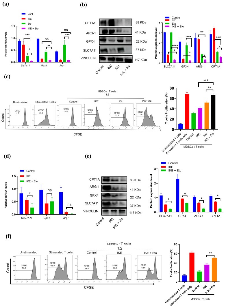
Figure 3.
IKE and Eto combined treatment blocked MDSCs’ immunosuppressive function by decreasing anti-ferroptotic and ARG1 proteins, thus promoting T-cell proliferation. The expressions of Slc7a11, Gpx4, and Arg-1 mRNA and proteins in in vitro BM-MDSCs (a,b) and in vivo spleen MDSCs (d,e) were detected by RT-qPCR or Western blotting, and protein expression levels were conducted by gray value analysis using ImageJ software (d,e). Percentages of T-cell proliferation in co-cultures of CFSE-labeled CD3+ T cells and in vitro BM-MDSCs (c) and in vivo spleen MDSCs (f) in a 2:1 ratio were analyzed by flow cytometry. Results from three independent experiments (a–f). Statistical analysis was performed using one-way ANOVA (a–f). Data are expressed as means ± SEM (n = 3). * p < 0.05, ** p < 0.01, *** p < 0.001, and **** p < 0.0001; ns, no significant difference.

