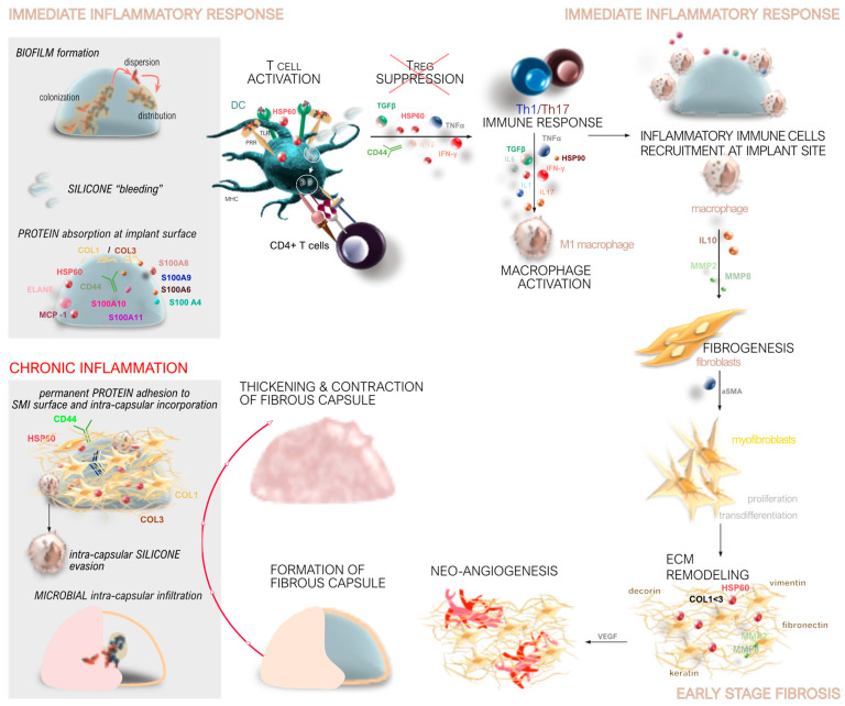
Figure 1.
Stages of capsular fibrosis around SMIs. Immediate wound response. During the acute wound healing phase, immediately after SMI implantation, the implant is exposed to wound bed fluid. T cells are activated primarily due to microbial contamination, implant shedding or silicone bleeding, and protein adsorption onto the implant’s surface and differentiate into Th1 and Th17 responses, while the T regulatory (Treg) response is suppressed. Foreign body response. Innate immune cells, such as neutrophils, macrophages, and dendritic cells, are recruited to the implant site. Macrophages are activated and play a key role in fibrogenesis, contributing to the early stages of fibrosis. Early-stage fibrosis The extracellular matrix undergoes remodeling with significant collagen deposition. The fibrotic tissue encapsulating the implant undergoes neo-angiogenesis, leading to the formation of a fibrous capsule. Chronic inflammation and capsular contracture. Chronic inflammation is perpetuated by the permanent adhesion of proteins to the SMI’s surface. Intracapsular silicone evasion and microbial infiltration further exacerbate the inflammatory response. This leads to excessive ECM remodeling and collagen deposition. The resulting thickened and contracted capsule causes discomfort and pain to the patient, potentially leading to implant displacement and necessitating revision surgery.

