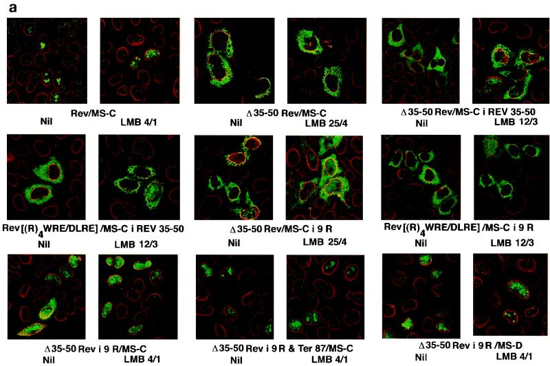
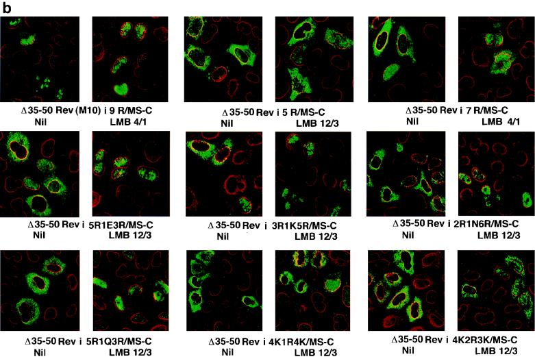
FIG. 4.
LMB-induced morphological changes in the subcellular localization of Rev and Rev–MS-C derivatives. (a) Changes in the distribution of mutants with deletions and insertions. (b) Changes in cells expressing proteins with multiple arginines or interrupted arginine or lysine strings. HeLa cells on 8-mm coverslips were transfected in quadruplicate with the indicated plasmids. Cells were rinsed and treated with LMB at 4 nM for 1 h (4/1), 12 nM for 3 h (12/3), or 25 nM for 4 h (25/4) or left untreated (Nil). At 24 to 36 h after transfection, transfectants were reacted with a mixture of rabbit anti-Rev antibodies and murine monoclonal antibodies against the nuclear pore complex. FITC-conjugated goat anti-rabbit IgG and Texas Red-tagged donkey anti-mouse IgG were used to label the fusion protein (green) and the nuclear pore complex (red). Images were visualized by using a ×63 objective lens on a Leica confocal microscope.


