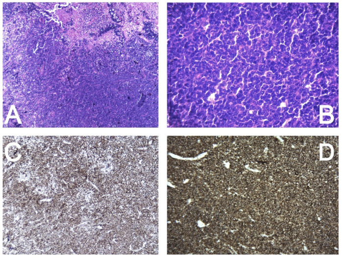
Figure 7.
Histopathology and immunohistochemistry report. Diffuse, with high cellular proliferation and extensive necrosis (4×—(A)). The tumor cells are large with irregular, atypical nuclei and distinct nucleoli, resembling centroblasts (40×—(B)). Ki67—diffusely positive in 90% of the tumor cells (C). CD20—strongly and diffusely positive in the tumor cells (D). CD34, CD10, and BCL6—negative expression in the tumor cells. MUM1—diffuse positive nuclear expression in the tumor cells. CD5—moderate positivity in most of the tumor proliferation. BCL2—diffusely positive in the tumor cells.

