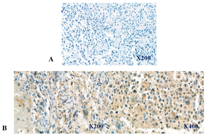
Figure 1.
Immunohistochemical expression of CD47 in biopsy samples. The depicted regions were selected from incisional biopsy samples, and immunohistochemical staining was performed as described under Materials and Methods. The upper panel (A, from case 14) depicts CD47-negative expression, while the lower panel (B, from case 15) depicts CD47-positive expression, showing CD47-positive cells stained in light brown color.

