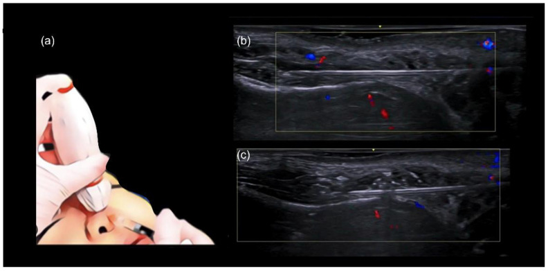
Figure 3.
Scan while injecting in the nose. (a–c) Color Doppler US with a 20 MHz probe demonstrates the in-plane technique in the dorsum, which allows real-time visualization of the cannula as filler is being deposited in a linear retrograde fashion. (a) Injection technique using a blunt cannula 25 G. Here we demonstrate the” scan while injecting” technique in the nose. (b) Plane of injection is checked for vessels. (c) Filler starts to be deposited.

