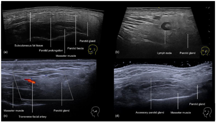
Figure 12.
US of the preauricular region layers. (a) Prominent anterior prolongation of the parotid gland. (b) A normal lymph node inside the parotid gland with a hyperechoic hilum (transverse panoramic and longitudinal view at 18 MHz, respectively). (c) Color Doppler highlighting the transverse facial artery emerging between the masseter muscle and the parotid gland. (d) Grayscale US demonstrating an accessory parotid gland superficial to the masseter muscle (transverse view at 18 MHz).

