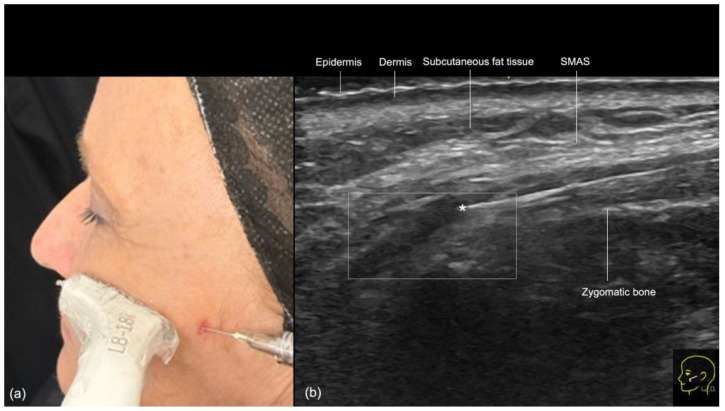
Figure 14.
Filler treatment of the supraperiosteal plane in the zygomatic region with a 22 G cannula. (a) Using the “scan while injecting” technique, the depth of the cannula can be controlled, ensuring that the supraperiosteal plane is reached. (b) Contact with vascular structures can be ruled out using color Doppler US imaging which shows the cannula in the supraperiosteal plane at 18 MHz and its tip (*) outside of vessels.

