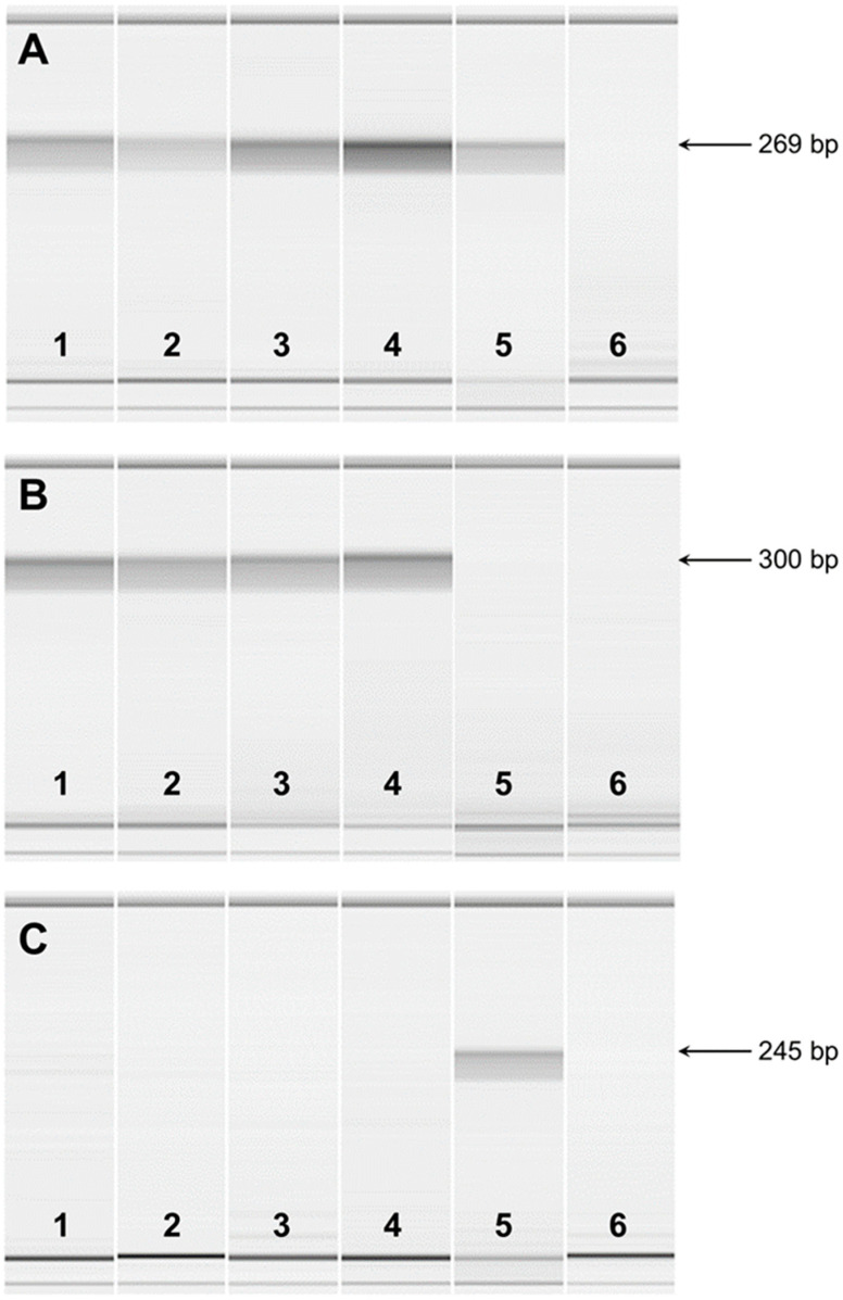
Figure 7.
Microcapillary electrophoretograms of RT-PCR amplicons from FAN1 mRNA. RT-PCR was performed with total RNA extracted from the kidneys of two FS-unaffected dogs (1 and 2), the blood of two FS-unaffected dogs (3 and 4), and one FS-affected dog (5). Lane 6 represents a negative control. (A) RT-PCR was performed with primers from exon 5 to exon 7 of the FAN1 gene. The expected amplicon size was 269 bp. (B) RT-PCR was performed with primers from exon 12 to exon 14 of the FAN1 gene. The expected amplicon size was 300 bp. (C) RT-PCR was performed with primers from exon 12 to intron 13 of the FAN1 gene. The expected amplicon size was 245 bp.

