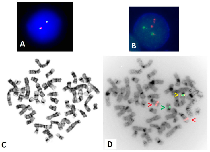
Figure 2.
FISH using the RARA break apart and PML/RARA dual fusion probes. (A) Interphase FISH for RARA break apart displaying a normal 2F pattern. (B) Interphase for PML/RARA dual fusion displaying an atypical abnormal pattern of 2R 1G 1F. (C) G-banded metaphase displaying a normal female karyotype. (D) Inverted DAPI image of metaphase PML/RARA FISH, with red arrows indicating PML on 15q24, a green arrow indicating RARA on 17q21, and a yellow arrow indicating a fusion. The PML::RARA gene fusion is present on a normal-appearing 17q.

