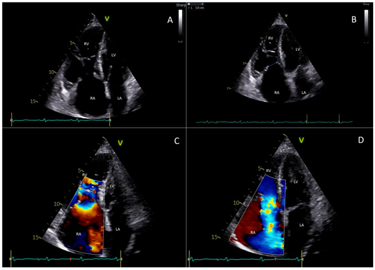
Figure 1.
This figure represents a case evaluated at Fondazione Policlinico Universitario Campus Bio-Medico, Rome. It is about a 77-year-old woman who suffers from carcinoid heart disease with involvement of the right heart chambers and tricuspid valve. (A) The transthoracic echocardiogram (4-chamber view) shows a significant dilatation of right heart chambers with “bulging” of the interventricular septum; the right papillary muscles, chordae tendineae, and tricuspid valve leaflets appear thickened; a diastolic restricted opening of the tricuspid valve is also appreciable. (B) The transthoracic echocardiogram (4-chamber view) in systole shows severe malcoaptation between the tricuspid septal and anterior leaflets creating a gap of 1.6 cm. (C) The color Doppler flow detects a stenotic filling in diastole. (D) The color Doppler in systole shows torrential tricuspid regurgitation. LA: left atrium; LV: left ventricle; RA: right atrium; RV: right ventricle.

