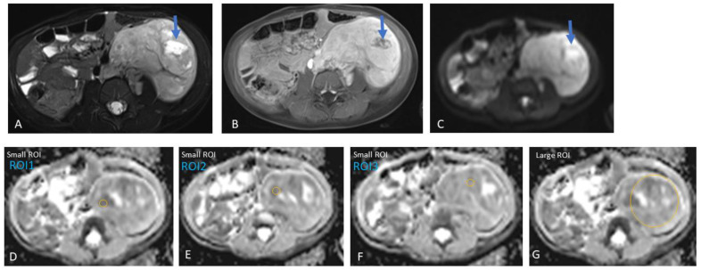
Figure 1.
Left retroperitoneal neuroblastoma with areas of necrosis in a three-year-old girl. Heterogeneous tumour, predominantly solid with a focal fluid area (arrow), potentially representing necrosis within the tumour. Therefore, this focal area of fluid was avoided during the placement of the regions-of-interest (ROIs) in the tumour for data analysis. (A) Axial T2, (B) axial post-gadolinium T1, and (C) axial DWI (b-value: 800 mm2/s). (D–F) Three small ROIs on the ADC map with an area of 19.1 mm2 and an average value of 1.23 × 10⁻³ mm2/s, and (G) one large ROI with an average area of 2721.1 mm2 and an average value of 1.42 × 10⁻³ mm2/s.

