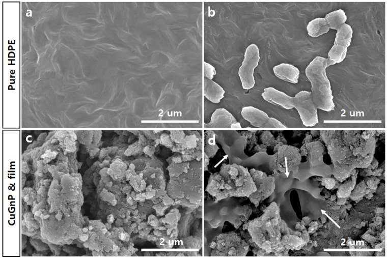
Figure 7.
Scanning electron microscopy (SEM) images of (a) high-density polyethylene (HDPE) film, (b) Escherichia coli on HDPE film, (c) copper-doped graphitic nanoplatelet (CuGnP) and HDPE film, and (d) E. coli on CuGnP and HDPE film after 6 h of culture. Arrows indicate membrane damaged E.coli. Scale bars represent 2 µm.

