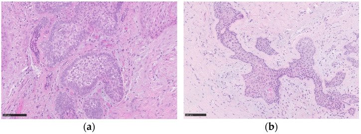
Figure 3.
Histopathological findings show (a) the lesion involving the oral stroma, consisting of epithelial islands characterized by peripheral basal cells with nuclear palisade and reverse polarization of the nuclei; the cells present in the center of the islands are loosely arranged and have a lighter cytoplasm than that of the cells of the basal layer. (b) In other deeper islands, the aspects described previously are less evident and below, in the center of an island, a focus of squamous differentiation is present. The black scale bars represent, in both the images, 100 µm.

