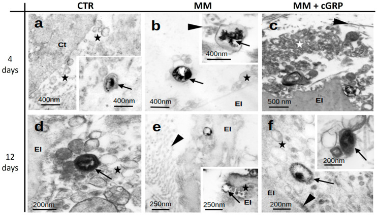
Figure 2.
Aortic tissue calcification is associated with calcified extracellular vesicles (EVs). Representative images of transmission electron microscopy (TEM) analysis of aortic tissues of Epon-Araldite resin ultrathin sections, cultured under control (CTR) (a,d), mineralizing (MM) (b,e), and MM supplemented with cGRP (MM + cGRP) (c,f) media for 4 and 12 days. Stars indicate extracellular vesicles; arrows indicate mineralized vesicles; arrowheads indicate collagen; El, elastin; Ct, cytoplasm of smooth muscle cell.

