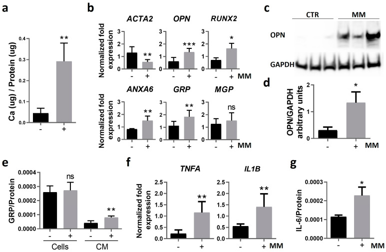
Figure 3.
Calcification of human primary VSMCs is characterized by osteoblastic differentiation and increased inflammation. Human primary VSMCs were cultured in control and mineralizing (MM) conditions for 14 days. (a) The mineralization rate was determined through calcium quantification normalized to protein levels. (b) Relative gene expression of VSMC osteogenic differentiation (ACTA2 encoding for ASMA, osteopontin (OPN), RUNX2, ANXA6) and mineralization inhibitor (GRP, MGP) markers by qPCR. (c) Western blot analysis for OPN detection and glyceraldehyde 3-phosphate dehydrogenase (GAPDH) as loading control. Original gels are presented in Figure S2; (d) relative OPN protein levels normalized to GAPDH using ImageJ software version V1.53f51 (arbitrary units). (e) Quantification of GRP normalized to total protein levels in cell lysates (cells) and in the cell culture media (CM) by ELISA. (f) Relative gene expression of VSMC proinflammatory cytokines (tumor necrosis factor alpha, TNFA; interleukin-1 beta, IL1B), and (g) quantification of interleukin-6 (IL-6) released to the cell media by ELISA, normalized to total protein levels. In all experiments, SD was calculated from 3 independent experiments (n = 3), and unpaired t tests were used. Statistical significance was defined as p ≤ 0.05 (*), p ≤ 0.01 (**), p ≤ 0.001 (***); ns, non-significant. In all graphs, black bars correspond to analysis at 4 days, and grey bars to 12 days.

