Figure 6.
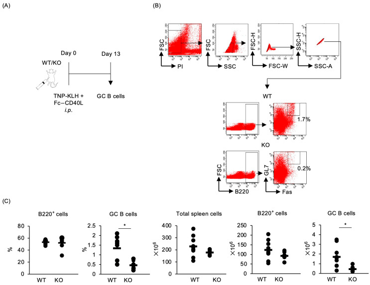
Traf5−/− mice primed with TNP-KLH and CD40L display defective development of GC B cells. The development of GC B cells in WT (n = 9) and KO (n = 7) mice was evaluated by flow cytometry. (A) Experimental schema. (B) Gating strategy for identifying PI-negative, Fas+GL7+B220+ GC B cells in the splenocytes of WT and KO mice immunized intraperitoneally with TNP-KLH and Fc-CD40L. (C) Percentages and numbers of total spleen cells, B220+ cells, and GC B cells in WT and KO mice, 13 days post-immunization. Bars represent average values for individual mice. * p < 0.05 (Student’s t-test).

