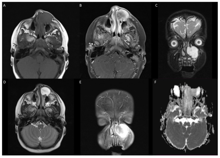
Figure 1.
(A) T1 weighted image showing hypointense lesion in left maxilla extending infraorbitally. (B,E) T1 with contrast showing avid heterogenous enhancement of lesion. (C) T2 coronal demonstrating superior and inferior extension of lesion. (D) T2 fat-suppressed axial with hyperintense lesion. (F) DWI showing no apparent hyperintensity or low ADC values.

