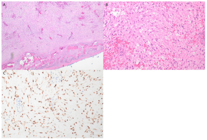
Figure 5.
(A) H&E ×4—low power image also showing loose myxoid stroma, although less prominent than with case 1; peripheral rim of reactive new bone was also seen focally around lesion. (B) H&E ×20—higher power showing spindle, stellate and some larger, more epithelioid cells, although without significant cytological atypia. (C) Beta-catenin ×20 demonstrates diffuse nuclear expression of tumor cells, noting that vessels within lesion are not stained.

