Figure 5.
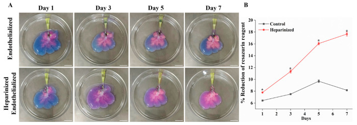
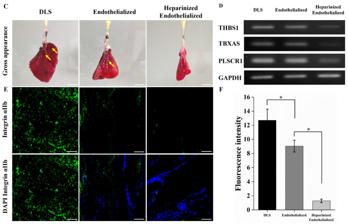
Resazurin reduction assay and ex vivo blood perfusion of re-endothelialized scaffold. (A) Resazurin reduction assay: visual photograph showing reduction of resazurin reagent from blue to pink over time, indicating cell proliferation in control and heparin-modified re-endothelialized liver scaffolds (Scale bar = 2 cm). (B) The curve shows significant proliferation of cells in heparinized re-endothelialized scaffolds compared to control (n = 3, * p < 0.05). (C) Gross appearance of heparinized re-endothelialized scaffold was free of clots compared to the non-coated re-endothelialized and DLS (yellow arrows indicate clot) (scale bar = 2 cm). (D) PCR analysis showed low expressions of thrombogenicity-related genes (THBS1; thrombospondin 1, TBXAS; thromboxane A synthase, PLSCR1; phospholipid scramblase 1) in blood-perfused heparinized re-endothelialized scaffolds compared to re-endothelialized and decellularized scaffolds. (E) Immunofluorescence staining with anti-integrin αIIb (green) and DAPI (blue) showing the platelets adherence and EC attachment in the scaffolds (scale bar = 100 μm). (F) Quantification of fluorescence intensity of integrin αIIb (green) demonstrates a significant reduction in intensity of heparin-treated re-endothelialized scaffolds, n = 4 fields, * p < 0.05.


