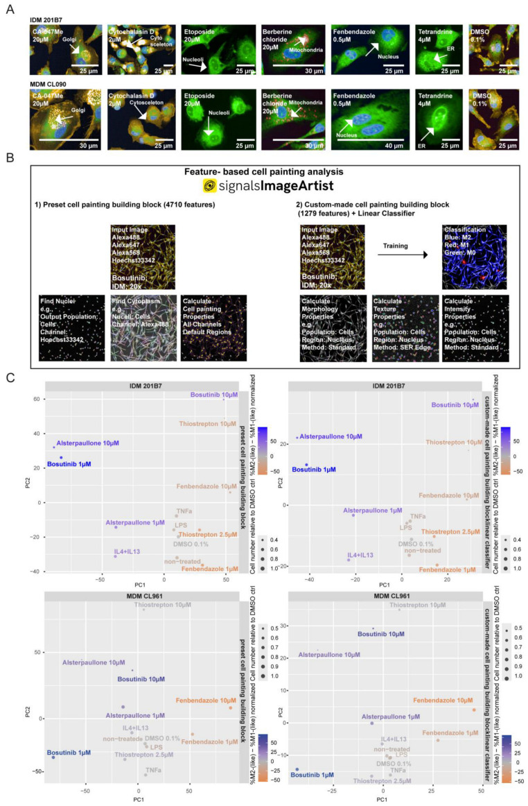
Figure 4.
Cell painting features for the description of the phenotypic spectrum of MΦ polarization. (A) Representative high-content Opera PhenixTM confocal images of cell-painted (PhenoVue Kit: PhenoVue 641 mitochondrial stain, PhenoVue Hoechst 33342 nuclear stain, PhenoVue Fluor 488-concanavalin A, PhenoVue 512 nucleic acid stain, PhenoVue Fluor 555-WGA and PhenoVue Fluor 568-Phalloidin) IDMs and an MDM donor CL090 with indicated cell painting controls (berberine chloride, fenbendazole, etoposide, cytochalasin D, CA-074Me and tetrandrine; 12 wells per condition; 10 fields per well; from two independent replicate 384-well plates) after 24 h of stimulation. The effect of the cpds on the respective cellular compartment and organelles is indicated by the white arrows. (B) Schematic of the established feature-based analysis using SImA: ‘preset cell painting building block’ and ‘custom-made cell painting building block combined with linear classifier’. bosutinib-treated IDM Opera PhenixTM confocal imagery is shown as a representative example. The whole analysis pipeline is described in Materials and Methods 4.8. (C) Principal component analysis of ‘Preset cell painting building block’ and ’Custom-made cell painting building block’ features indicated cpd- and biologically stimulated IDMs and an MDM donor CL961 for 24 h. Each datapoint indicates the mean of 4 replicate wells (5 fields per well; 20× magnification) per condition. ‘Preset cell painting building block’ is based on 4710 used features. ’Custom-made cell painting building block’ is based on 1279 features.

