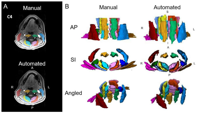
Figure 1.
(A) Axial cervical spine muscle segmentations at the C4 vertebral level from manual segmentation and an automated computer-vision model overlaid over a water image from Dixon fat–water MRI. (B) Three-dimensional renderings of cervical spine muscle segmentations. The muscle groups segmented include the multifidus and semispinalis cervicis (left = light pink, right = aqua), longus colli and longus capitis (left = light green, right = gold), semispinalis capitis (left = orange, right = yellow), splenius capitis (left = dark pink, right = light blue), levator scapula (left = indigo, right = dark green), sternocleidomastoid (left = blue, right = red), and trapezius (left = brown, right = magenta). L = left, R = right, A = anterior, P = posterior, S = superior, I = inferior. Adapted from Weber et al., 2021 [5].

