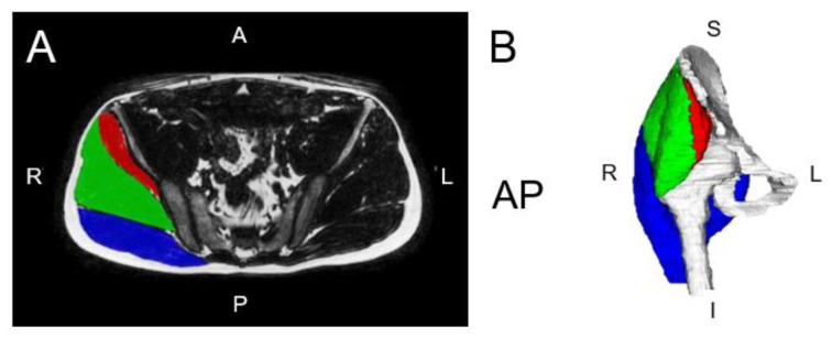
Figure 3.
(A) Axial right hip muscle segmentation overlaid over a fat image from Dixon fat–water MRI. (B) Three-dimensional renderings of the hip muscle segmentations. The muscle groups segmented include the gluteus maximus (blue), gluteus medius (green), and gluteus minimus (red). Adapted from Perraton et al., 2024 [69].

