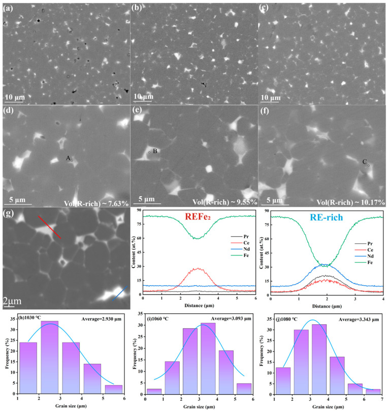
Figure 3.
Backscattered electron scanning electron microscopy (SEM) images of sintered magnets at 1030 °C (a,d), 1060 °C (b,e), and 1080 °C (c,f). (g) Scanning magnification of the magnets at 1080 °C and line scans of the REFe2 phase and the RE-rich phase, as well as grain sizes of the sintered magnets at 1030 °C (h), 1060 °C (i), and 1080 °C (j).

