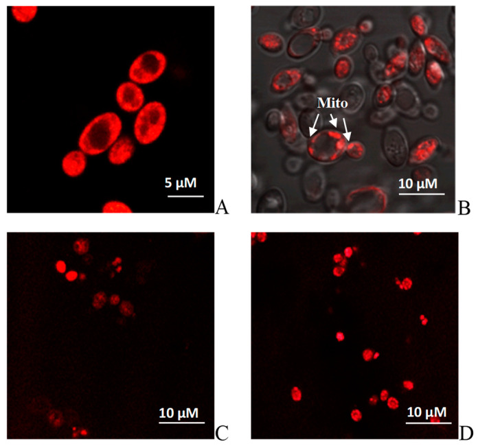
Figure 3.
Potential-dependent staining of the mitochondria in Y. lipolytica W29. (A,B)—pH 5.5; (C)—pH 4.0; (D)—pH 9.0. Cells were incubated with 0.5 µM MitoTracker Red for 20 min. The incubation medium contained 0.01 M PBS, 1% glycerol, pH 7.4. The areas of high mitochondrial polarization are indicated by bright-red fluorescence due to the concentrated dye. To examine the MitoTracker Red stained preparations, filters 02 and 15 (Zeiss) were used (magnification 100×). Photos were captured using an AxioCam MRc camera.

