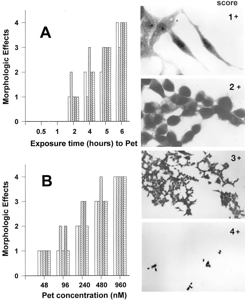
FIG. 2.
Quantitation of time- and dose-dependent effects of Pet protein on HEp-2 cells. (A) Time-dependent effects of Pet protein. The effects of Pet protein on the morphology of HEp-2 cells were examined after 0.5, 1, 2, 4, 5, and 6 h of exposure. After this time, monolayers were scored as described in the text. The bars represent individual experiments (n = 4 for each time point). (B) Dose-dependent effects of Pet protein. HEp-2 cells were exposed to different concentrations of Pet protein (48 to 960 nM). The bars represent individual experiments (n = 4 for each concentration point). The inserts show the score of the morphologic changes of HEp-2 cells produced by Pet protein described in Materials and Methods.

