FIG. 1.
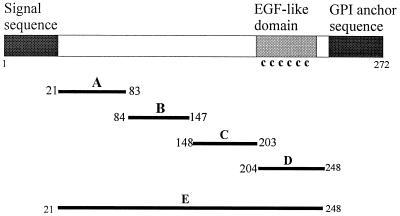
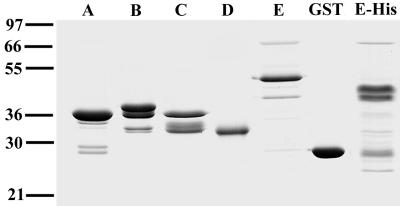
(Top) Structure of MSP4 and positions of the expression constructs. Black boxes at the left and right represent the signal sequence and the GPI anchor sequence, respectively; the shaded box indicates the EGF-like domain which contains the six cysteine residues. A, B, C, and D are four MSP4 fragments cloned in pGEX vectors, and E is the full-length MSP4 lacking signal and anchor sequences cloned in both pGEX and pTrcHis vectors. The first and last amino acid residues of MSP4 included in each recombinant protein are indicated. (Bottom) Coomassie blue-stained SDS-PAGE gel showing the five recombinant MSP4 GST fusion proteins GST-MSP4A (lane A), GST-MSP4B (lane B), GST-MSP4C (lane C), GST-MSP4D (lane D), and GST-MSP4E (lane E) and the hexahistidine fusion of full-length MSP4 (lane E-His). As a control, GST is included. Positions of molecular mass standards (kilodaltons) are shown at the left.


