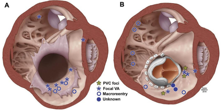
Figure 3.
This illustration depicts the right ventricle in a typical patient with Ebstein’s anomaly prior to (A) and post-TV replacement (B). The axial section of the heart is visualized in a left anterior oblique view, from the ventricle, below the valve. The most common site of origin for ventricular arrhythmias in unoperated patients was in the atrialized right ventricle. Post TV replacement, the sites of origin (focal) and slow zones (macroreentry) of the ventricular arrhythmias were diverse [32].

