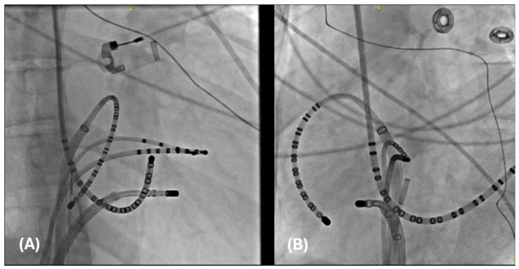
Figure 4.
Catheter placement of catheters in preparation for an invasive electrophysiological study. (A) A right anterior oblique (RAO) image of a duodecapolar Cristacath catheter (A20) catheter inside an SR0 sheath along the tricuspid annulus, another duodecapolar catheter placed in the coronary sinus, a quadripolar catheter advanced to the RV apex, an octapolar catheter advanced to the His position, and an ablation catheter within a steerable sheath positioned in the anterolateral tricuspid annulus. (B) The same catheters are pictured in the left anterior oblique (LAO) view.

