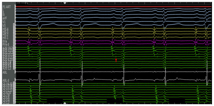
Figure 5.
Electrograms of the aforementioned 27F with Ebstein’s anomaly in sinus rhythm. Catheters used and their respective positions are indicated in Figure 4. The surface ECG and intracardiac EGMs are labeled on the left side of the figure. Normal AH and HV intervals are present at the baseline with no manifest pre-excitation. There is a near-field potential on the A20 catheter along the lateral tricuspid annulus (red arrow). This precedes the His signal and is most consistent with a pathway potential.

