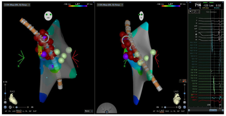
Figure 9.
Electroanatomic map showing an RAO (left) and LAO (middle) view of the A20 catheter and radiofrequency ablation lesions in the lateral tricuspid annulus corresponding to the EGM of the pathway potential and earliest point of activation (right). Catheter positions were previously described in Figure 4. RAO = right anterior oblique. LAO = left anterior oblique. EGM = electrogram.

