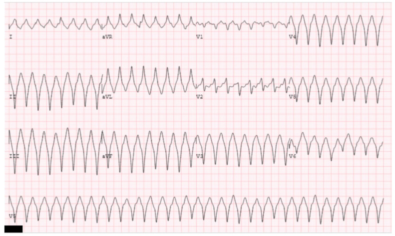
Figure 10.
The clinical ventricular tachycardia is illustrated. ECG morphology of left bundle, left, superior axis suggests a ventricular tachycardia originating from the basal, inferior wall of the right ventricle. Note there is a reverse pattern break likely indicating exit near the basal septum [37].

