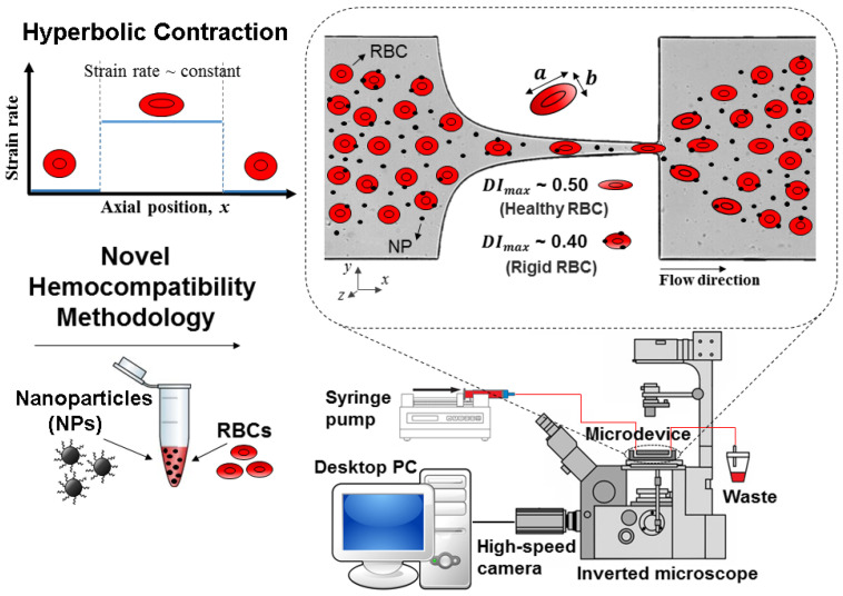
Figure 8.
Representation of the procedure for evaluating the hemocompatibility of RBCs in contact with MNPs. The a and b represent the major and minor lengths of the ellipse, respectively. A high-speed video microscopy system and a PDMS microfluidic device with a hyperbolic constriction microchannel make up the microfluidic methodology, adapted from [208].

