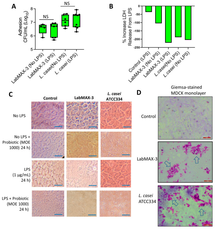
Figure 5.
Adhesion characteristics of LabMAX-3 in the MDCK cell line. (A) Adhesion (CFU/mL) of probiotic cultures to MDCK cells. (B) Lactate dehydrogenase (LDH) release assay from MDCK cells during the LabMAX-3 adhesion experiment. (C) Light microscopic analysis of cell monolayer integrity during probiotic adhesion experiment. Scale: 25 µm. (D) Giemsa staining of MDCK cells after probiotic exposure. LabMAX-3 forms biofilm-like structures in patches (arrows) on MDCK cell monolayers. Scale: 50 µm.; NS, not significant.

