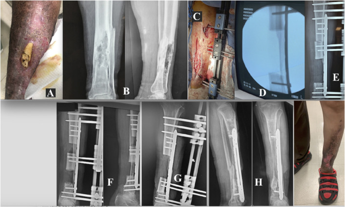
Fig. 10.
Figs. 10-A through 10-H Patient 2. Fig. 10-A Osteomyelitis in the distal third of the left tibia. Fig. 10-B The involved bone segment. Fig. 10-C Placement of the MIRP. Fig. 10-D Resection of infected and necrotic bone tissue. Fig. 10-E Initial compression of 3.5 Nm. Fig. 10-F The MIRP rail is used for transport of the bone to its final destination and control of its docking. Fig. 10-G Completion of bone transport, with a final compression of 3.1 Nm. Fig. 10-H Complete consolidation has resulted in functional, uninfected bone.

