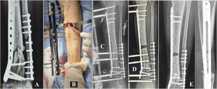
Fig. 4.
Figs. 4-A through 4-E Patient 15. Fig. 4-A Infected nonunion of the distal third of the left tibia. A previous osteosynthesis attempt failed, with 24° varus deviation of the distal fragment and joint. Fig. 4-B Removal of osteosynthesis material and placement of the MIRP (minimally invasive technique) and the initiation of osteotomy. Fig. 4-C Immediate correction of the tibial and joint axes. Fig. 4-D Control of docking by means of the MIRP rail, with final compression on the infected PN. Fig. 4-E Removal of the device and docking site consolidation healing of the infected PN (4.4-Nm final compression), and formation of osseous trabeculation in the distraction regeneration zone.

