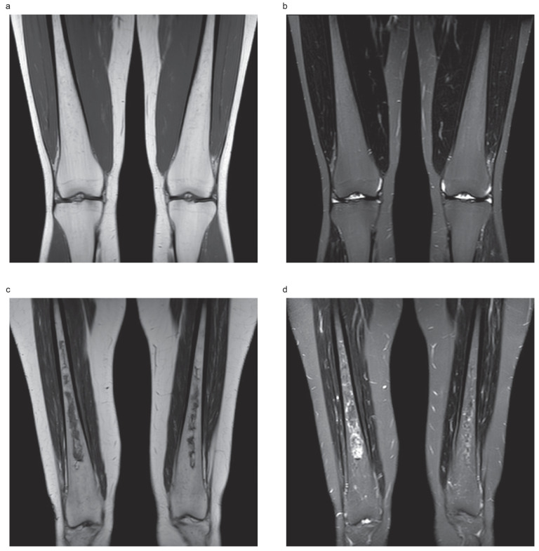
Figure 1.
Typical appearance of heterogenous Gaucher marrow fat signals with no evidence of osteonecrosis. (a) Coronal T1-weighted image shows several punctate low-signal foci scattered throughout the diaphyseal regions of both femora, corresponding to infiltration of the marrow by Gaucher cells in a 35-year-old woman with Gaucher disease type 1. (b) The corresponding coronal Short Tau Inversion Recovery (STIR) image. Typical appearance of an established osteonecrosis. (c) Coronal T1-weighted image shows irregularly bordered areas of hypointensity within the diaphyseal regions of both femora, corresponding to fibrosed and sclerosed bone marrow of established bone infarcts in a 50-year-old woman with Gaucher disease type 1. (d) The same areas show serpiginous inner rim of hyperintensity on the corresponding coronal Short Tau Inversion Recovery (STIR).

