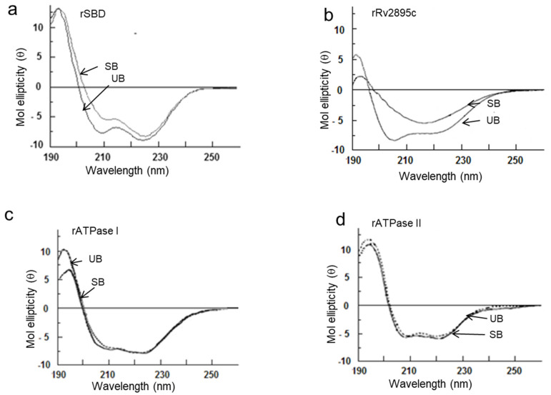
Figure 4.
Only rSBD and rRv2895c show substrate-induced secondary structure in far-UV CD spectra, indicating substrate-induced secondary structural changes. CD analyses of (a) rSBD, (b) rRv2895c, (c) rATPase I, and (d) rATPAse II were carried out in unbound (UB) and substrate-bound (SB) forms. The spectra shown here are resultant after normalizing with buffer and ligand spectra. Five readings were taken in each case.

