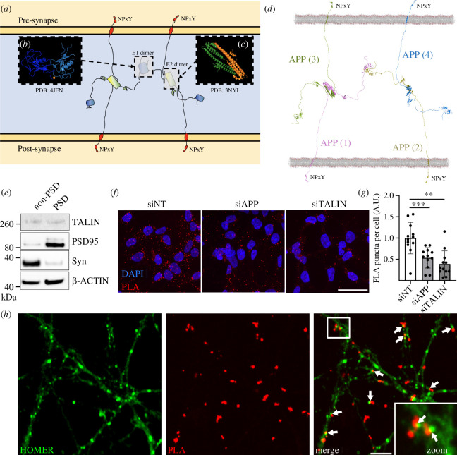
Figure 3.
APP dimerization leads to the formation of an extracellular synaptic meshwork. (a) Four APP molecules are shown forming a dimer of dimers. (b, c) The crystal structures of (b) the E1 dimer [50] and (c) the E2 dimer [51]. These dimer interfaces were used to overlay four full-length APP structural models. (d) Structural model of the APP synaptic meshwork. The four APP molecules are numbered, 1–4, with APP molecules 1 (pink) and 2 (yellow) embedded in the post-synaptic membrane, and 3 (green) and 4 (blue) embedded in the pre-synaptic membrane. On the cytoplasmic face of both synaptic compartments are positioned NPxY motifs that are spatially organized by the APP oligomerization. See also electronic supplementary material, video 2. (e) Synaptic fractionation experiment revealed the presence of talin1 in both pre- and post-synaptic compartments. Post-synaptic density (PSD). (f–h) Proximity ligation assay (PLA) of APP and talin1 in cells and neurons. (f) PLA of APP/talin (red) signal in HEK293-APP695WT transfected with siRNAs targetting APP or talin. A non-targetting (NT) siRNA was used as a control. The nucleus is visualized using Hoechst (blue). Scale bar = 40 µm. (g) Quantification of PLA as performed in (f). (h) APP/talin PLA puncta (red) were also observed at synapses from primary neuronal cultures. Homer staining (green) was used as a synaptic marker. Arrows show the proximity between PLA signal and synaptic marker. Scale bar = 4 µm. **p < 0.01,***p < 0.001, Mann–Whitney non-parametric test.

