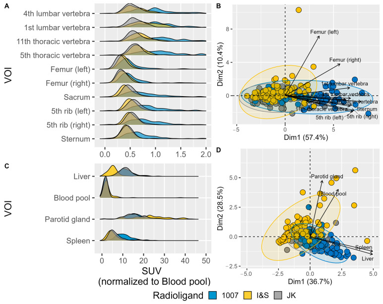
Figure 4.
Normalized SUV peak values in different Volumes of Interest (VOIs). (A) Density plots showing the distribution of normalized SUV values for various bone VOIs. (B) PCA biplot visualizing the relationships among skeletal VOIs based on their peak SUV values. Different colors indicate the three radioligands, and arrows show the direction and magnitude of the VOI contributions to the principal components. The length of each vector represents the importance of the corresponding VOI in the PCA, with longer vectors indicating a greater contribution to the variance. The direction of the vectors indicates how each VOI is related to the principal components, with similar directions suggesting similar patterns of variation. (C) Density plots showing the distribution of peak SUV values (g/mL) for various reference VOIs. (D) PCA biplot visualizing the relationships among reference VOIs based on their peak SUV values. (The more readable (B) panel can be found as Figure A2).

