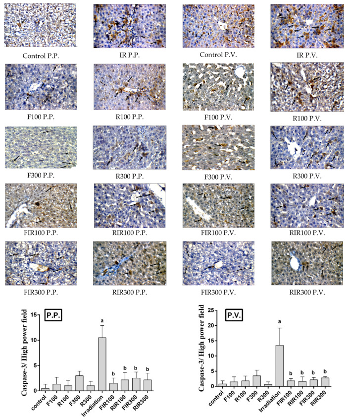
Figure 8.
Immunohistochemical photographs of caspase-3 expression in hepatic peri-portal (P.P.) and peri-venular (P.V.) (black arrows) areas in irradiated rats treated with W. filifera and W. robusta leaves ethanolic extracts (×400). Sections taken from livers (P.P. and P.V.) of control rats showing minimal expression. Sections taken from livers (P.P. and P.V.) of irradiated rats shows extensive cytoplasmic expression (brown color). Sections taken from livers (P.P. and P.V.) of irradiated rats treated with W. filifera or W. robusta showing medium to limited expression (brown color). Data conveyed as mean ± SD (n = 6), significance was at p ≤ 0.05 by means of one-way ANOVA followed by Tukey–Kramer as a post hoc-test. a Significantly different as of control group. b Significantly different as of irradiated group.

