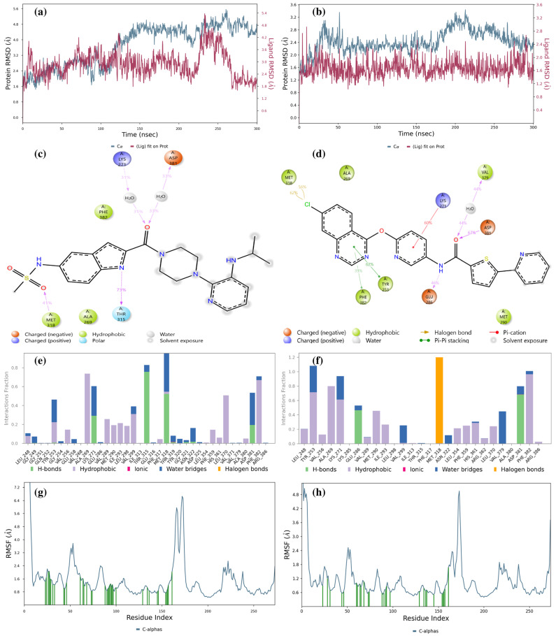
Figure 8.
Result of 300 ns MD analysis for LMQC01 and LMQC04 binding to the Abl-kinase domain. The protein–ligand RMSD plot of LMQC01 (a) and LMQC04 (b) bound to the Abl-kinase domain (PDB ID: 1IEP). Ligand–protein contact interactions scheme with the protein residues of LMQC01 (c) and LMQC04 (d) bound to Abl-kinase. Protein–ligand contacts histogram of the interaction fraction of H-bond (green), hydrophobic bond (purple), ionic bond (magenta), water bridges (blue), and halogen bonds (orange) for LMQC01 (e) and LMQC04 (f). RMSF plot of LMQC01 (g) and LMQC04 (h) protein–ligand complex.

