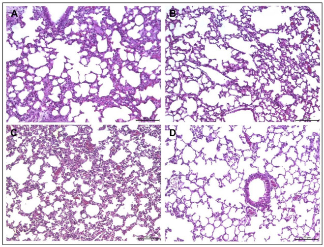
Figure 7.
Lung histology of IFN-y KO mice infected with M. abscessus or M. massiliense and treated with Agelaia-12. M. abscessus (A) or M. massiliense (C) infected animals presented difuse inflammatory lesions. After treatment with Agelaia-12, mice infected with M. abscessus (B) showed visually less inflammatory lesions than nontreated mice. Siimilarly, mice infected with M. massiliense and treated with Agelaia-12 showed visually less inflammatory lesions (D) than nontreated mice.

