Figure 1.
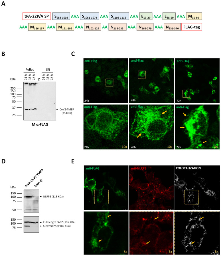
In vitro characterization of a DNA vector expressing the multiepitopic CoV2-TMEP protein. (A) Schematic representation of the chimeric CoV2-TMEP multiepitopic vaccine targeting T cells. (B) Time-course expression of CoV2-TMEP protein. HEK-293T cells were transfected as detailed in the Materials and Methods section and CoV2-TMEP expression in pellets and supernatants (SN) was analyzed by Western blotting using the mouse monoclonal anti-FLAG antibody. (C) Subcellular localization of CoV2-TMEP protein. HeLa cells were transfected and processed for immunofluorescence microscopy as indicated in the Materials and Methods section. Green staining: mouse monoclonal anti-FLAG antibody followed by anti-mouse Alexa Fluor 488 antibody. Yellow arrows: CoV2-TMEP forming aggregates in magnified images (10×). (D) Analysis of NLRP3 induction and PARP cleavage. HEK-293T cells were transfected as described in the Materials and Methods section and the expression of NLRP3 (118 KDa; upper panel) and PARP (full-length: 116 KDa; cleaved: 89 KDa; lower panel) was analyzed by Western blotting using specific antibodies. (E) Subcellular colocalization of CoV2-TMEP protein with inflammasome. HeLa cells were transfected and processed for confocal microscopy as detailed in the Materials and Methods section. A mouse monoclonal anti-FLAG antibody and a rabbit polyclonal anti-NLRP3/NALP3 antibody followed by the corresponding secondary antibodies conjugated with Alexa Fluor 488 (green staining; FLAG detection) or Alexa Fluor 594 (red staining; NLRP3 detection) were used. Colocalization signal is indicated in white. Yellow arrows: Discrete colocalization signals in magnified images (5×). One representative optical section from three independent experiments is shown.

