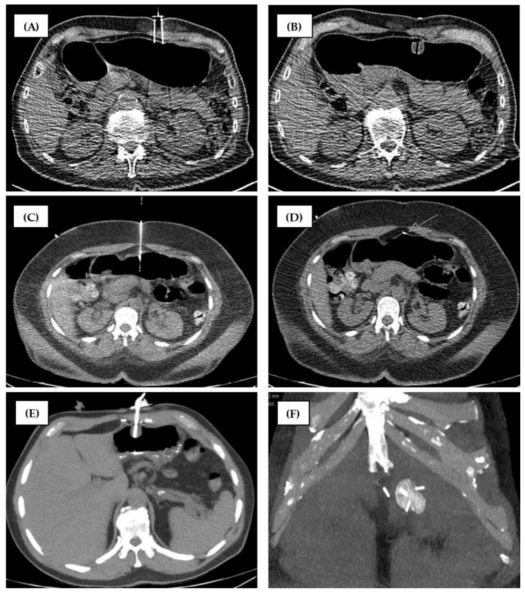
Figure 4.
Comparison of fixation techniques. (A,B) technique of center 2. (C–F) technique of center 1. (A) Gastropexy Device II (Fresenius Kabi, Bad Homburg vor der Höhe, Germany) with two parallel hollow needles and a thread inside. (B) After gastropexy, the suture is not visible on CT. (C) A first SAF-T-PEXY T-Fastener anchor (Avanos Medical, GA, USA) was placed in the body of the stomach. (D) The anchor is visible in the CT. The arrow shows the correct end position on the inner stomach wall. (E) Gastric tube placed between the anchors. (F) The distances between the anchors are different. As usual, the tube was placed in the center of the triangle.

