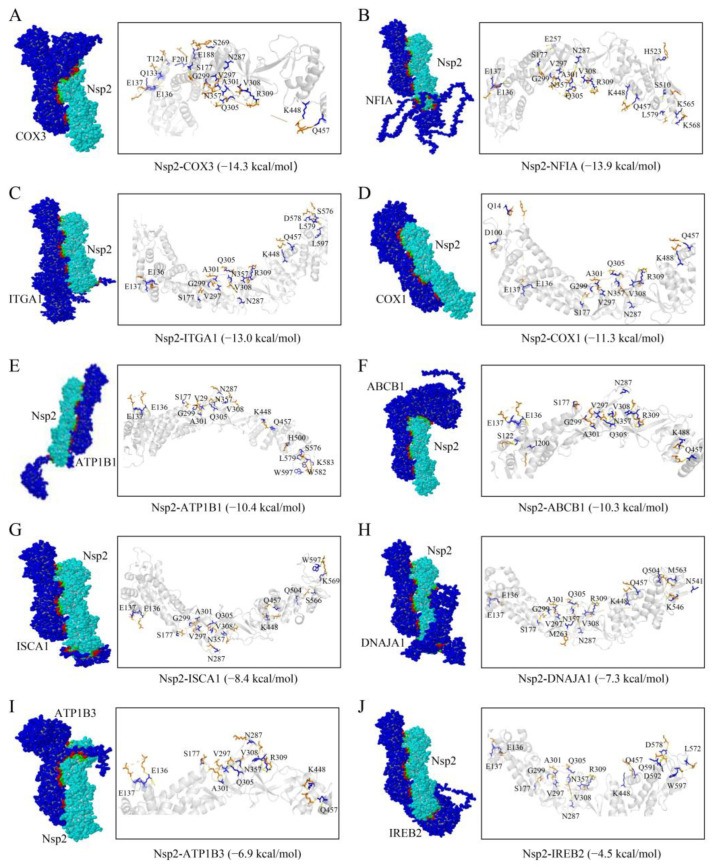
Figure 3.
Diagram of the binding pattern of the Nsp2 protein to the host proteins (A) COX3, (B) NFIA, (C) ITGA1, (D) COX1, (E) ATP1B1, (F) ABCB1, (G) ISCA1, (H) DNAJA1, (I) ATP1B3, and (J) IREB2. The interaction residues of the Nsp2 protein with the host proteins are shown as blue sticks, while the host proteins are denoted by orange sticks.

